Another project
Project Title.
Validation study of 3D printing prototypes of the temporal bone for surgical simulation (de vuelta)
Prototyp made in Alemania
Project Summary
Learning the art of surgery, and when it comes to ear surgery, requires rigorous training. The temporal bones of cadavers are ideal for this purpose, although currently the acquisition of these is difficult. On the other hand, technological advances, specifically in regards to 3D printing offer new opportunities. Achieving excellence in print quality and exact resemblance to real bone apparently requires expensive equipment. Based on a study carried out by medical students in 2019 where they carried out a review to evaluate the feasibility, in our environment, of access to 3D printing of the temporal bone, it was possible to know about the existence of several national specialized centers that could offer the necessary technology to achieve interesting results (1). Based on the above, we intend with this work to evaluate the quality of the 3D impression of the temporal bones of our patients, using different technologies available in our environment.
Research team
Area and specialty of the proposal
Medicine area in the specialty of otorhinolaryngology and based on digital manufacturing
Project objectives
Major:
Evaluate the quality of 3D printing of the temporal bones of our patients, using different technologies available in our environment.
Secondaries:
-
Update existing centers with 3D printing technology in our environment
-
Compare the technology, material used and cost with the quality of 3D printing
-
Define the path to follow in order to access excellent technology
Expected results
Gain a scale-based appreciation of the appearance, feel of consistency, and anatomical landmarks of different temporal bone 3D impression prototypes.
Background and Justification
3D printing of the temporal bone (HT) has been anatomically validated when compared to tomographic images and has additionally improved drilling accuracy and spatial representation during mastoidectomy simulation. (2.5)
There are several published works that compared the different types of materials used and different models of machines that evaluate the similarity with real bone and describe interesting results that encourage the use of this technology (2,3), other authors try to find a low-cost technology. cost and that presents valid results for use in surgical training (4)
Due to the lack of national data regarding 3D printing of the temporal bone and the need for a training method to develop surgical skills in otological surgery, we consider it important to carry out this study. Therefore, we propose to carry out an experimental work with 3D printed pieces of temporal bones using different types of technology and materials existing in our environment with the aim of determining which of the technologies and/or materials are closest to real bone.
Materials and Methods
Prospective study
A total of five participants included three third-year residents, a second-year Ear Fellow, and an otologist experienced in otologic surgery. Residents will be assisted by a more experienced surgeon throughout the drilling process.
Excluded participants: those with no experience in ear drilling
All participants will have a real temporal bone to closely experience possible differences with the prototypes and a number to be defined, probably 5 samples based on experiences from previous jobs (2), of prototypes manufactured with different printing instruments and different materials.
Evaluation of prototypes
All participants will have a list of the variables that they must take into account during milling Variable list adapted from a previous work (2):
- 1- Appearance of the prototype: 0 - 10
- 2- Milling sensation: 0 – 10
- 3- Preservation of anatomy: 0 -10
Prototype Appearance:
Please rate the relative appearance of the prototype compared to real bone, on a scale of 0-10 where 0 being not at all representative and 10 being perfectly representative
Milling feel:
Please rate the milling feel of the prototype compared to real bone, on a scale of 0-10 where 0 being not at all representative and 10 being perfectly representative
Anatomy Preservation:
Subitems to evaluate:
mastoid cells
sigmoid sinus
sinusoidal angle
Joint
Horizontal semicircular canal
Facial nerve
ossicular chain
Please rate on a scale of 0-10 where 0 being not at all representative and 10 being perfectly representative
The participants will have cutting and diamond burs of various sizes and a “Storz” brand motor used in the dissection laboratory.
Based on the investigation of the existing national 3D printing centers in our environment, the 3D printing machines will be defined, as well as the different types of materials.
To calculate the results, an average (mean) of the scores obtained will be obtained.
Prototype development:
The DICOMs that we will use will correspond to our patients and must be thin sections of at least 0.5 mm.
For the 3D reconstruction we will use the 3D_slicer software because it is a free access program and of very good quality. We will also rely on 3D_builder with which the HT cuts will be made to print each portion separately and then assemble all the portions.
This will allow us to remove possible internal supports that do not belong to the original structure (5).
STL 3D printing
Once the model to be printed is ready, it will be printed in the different selected equipment with the respective material.
Relevance of the proposal
It is a project that aims to explore new horizons with regard to the development of skills in the technical aspect, betting on technology and innovation.
Due to the lack of national data regarding 3D printing of the temporal bone and the need for a training method to develop surgical skills in otological surgery, we consider it important to carry out this study.
Plan of work
Duration time in months: 6 months Start of the project: July / 2022 Project Completion: December /2022
Work scheme

budget

Communication strategy
Once we have obtained results, these will be disseminated through a publication in an indexed journal to achieve greater reach.
Bibliographic references
- Gaona Rivas EP; Rojas Galeano WO; Romero Riveros MM; Ruiz Cano VR; Samaniego Rojas GI; Samudio Duarte TA; Roig Ocampos Jr. Reconstruction of the temporal bone by 3D printing as a learning method. Monograph. Chair of Otorhinolaryngology UNA. 2019
- Max Haffner, BS, Austin Quinn, BA; Tsung-yen Hsieh, MD, E Bradley Strong, MD; Toby Steele, M.D. Optimization of 3D prompt material for the recreation of patient-specific temporal bone model. Annals of Otology, Rhinology & laryngology 127(5), 338-342, 2018.
- Horcacitas-Pous R; Gonzalez A, The importance of creating a model of the temporal bone in a 3D printer for the milling laboratory. Review of recreated cases. An Orl Mex 65 (4): 205-211, 2020;
- Mowry SE; Jammal H; Myer Ch; Solar AC; Weinberger P. A novel temporal bone simulation model using 3D printing techniques. Otology & Neurotology 36: 1562-1565, 2015
- Hochman JB, Kraut J, Kazmerik K, Unger Bertram J. Generation of a 3D printed temporal bone model with internal fidelity and validation of the mechanical construct. Otolaryngology Head and Neck Surg 150: 448-54, 2014.
Experience outside of proyect
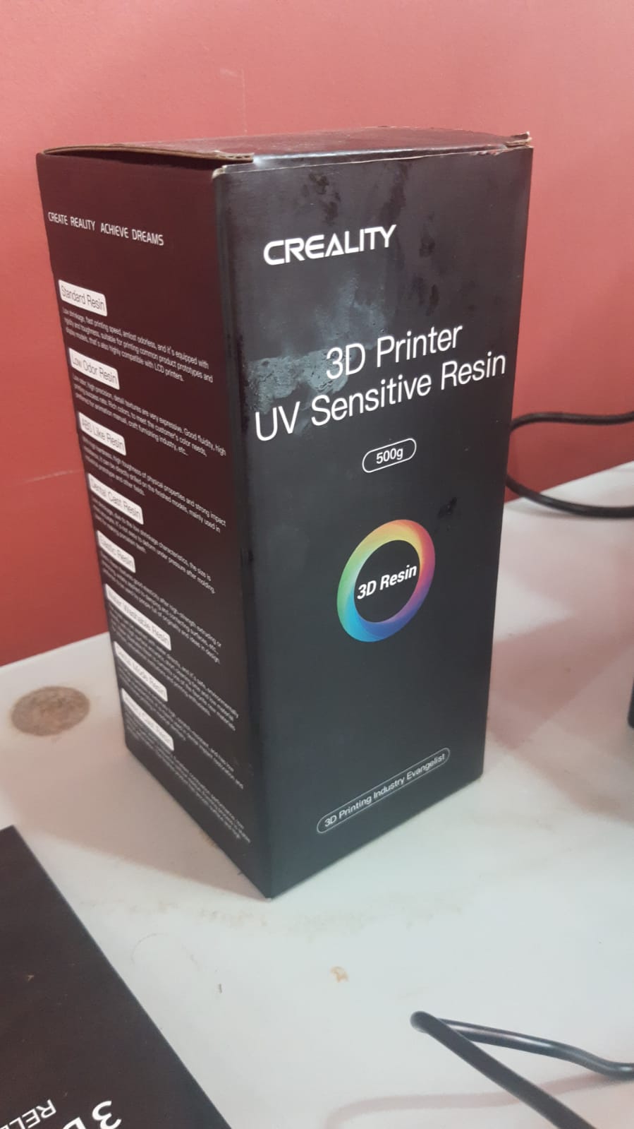
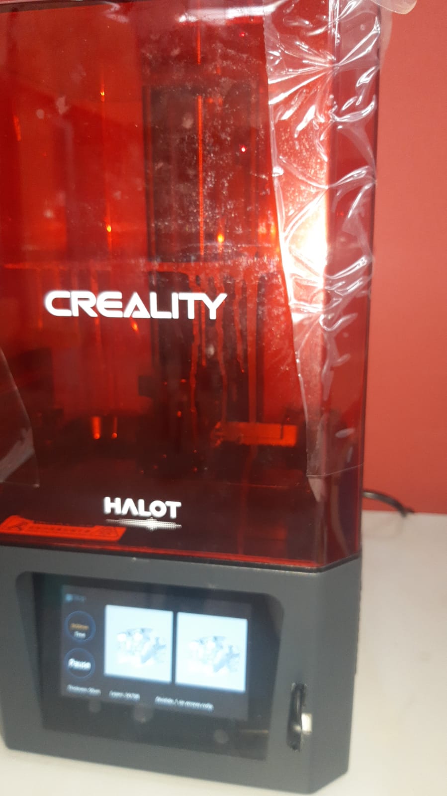
This prototype was made with Creality Printer, this work with resina and ultraviolet curing
First experience whit incolor resin
Set of photos but of the entire temporal bone
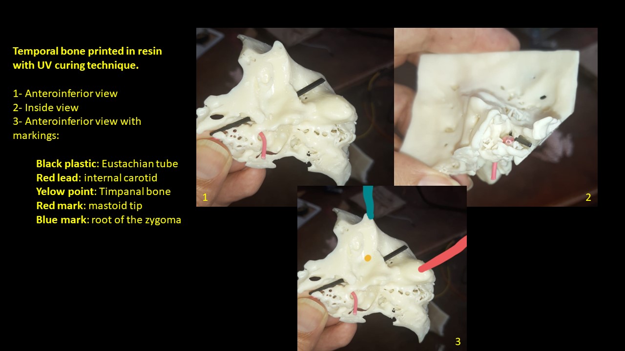
Second experience
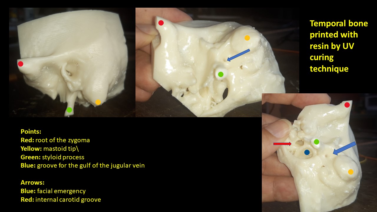
I am experimenting with the [3D Slicer] (https://www.youtube.com/watch?v=Dn3ANveKQXY) software and edited the temporal bone of a patient who has already undergone surgery with a diagnosis of carcinoma of the external ear canal
It was a very laborious surgery and consisted of removing the tympanal bone avoiding noble structures such as the facial nerve, large vessels (gulf of the jugular) and above the meninges.
When doing the 3D impression I could see the close relationship with the carotid artery.
We just had difficulty removing the piece despite the fact that it seemed to be already loose and we had to refine a part of the tympanal bone on its anterior face that extended towards the bottom and in this way with a little skill we got it.
Precisely that bone remnant that remained had a very close relationship with the internal carotid artery.
Resin Models
Light induces the polymerization of multifunctional monomers; it is what we know as ‘UV light curing’ and it is recognized as the most effective system to transform very quickly and at room temperature a solvent-free liquid resin into a highly resistant polymeric product.
This is the patient’s temporal bone still with the impression supports
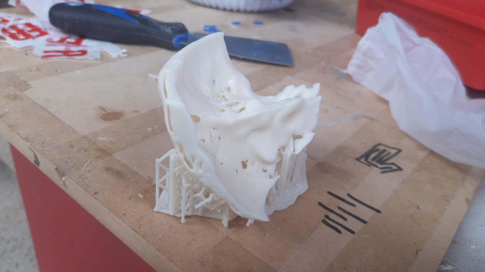
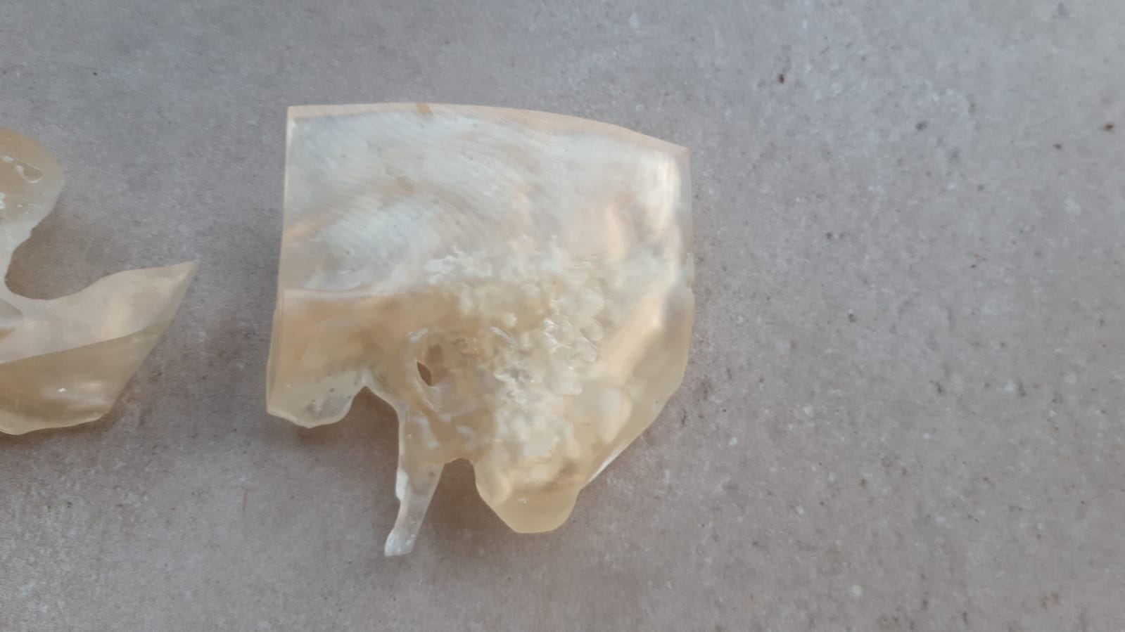
This is another temporal bone, the same as the first photos (first experience) but this time I printed it in its entirety without dividing it like the previous one, also still with the supports
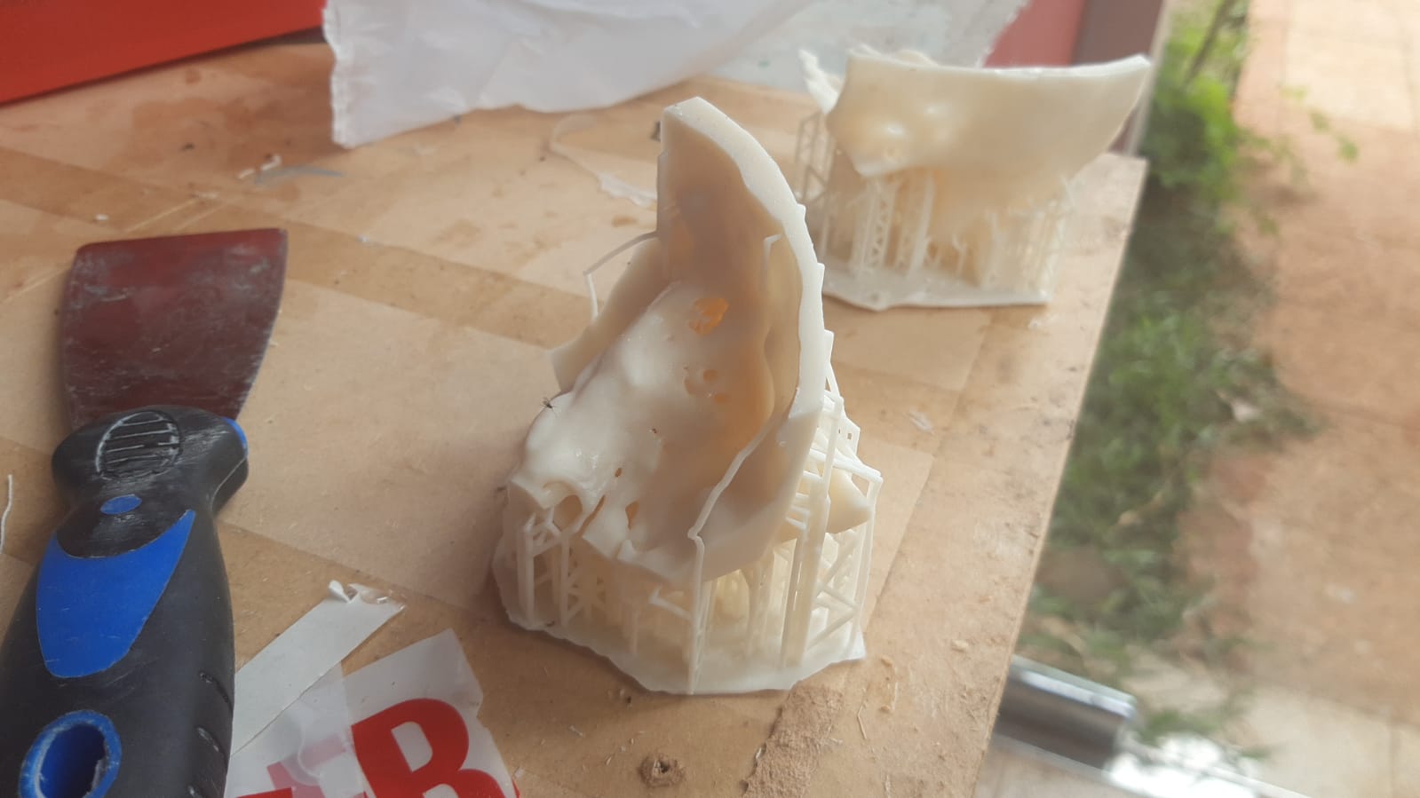
Set of photos of temporal bone 3D printing of the Second Experience
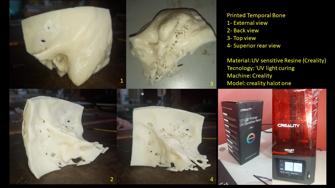
Photo of the Second Experience Bone showing the relationships of the temporal bone with the great vessels. An anterior and inferior view of the same
Continuing with my project
My first experience of 3D printed temporal milling
In the temporal bone dissection laboratory of the ENT Service of the UNA Medical Sciences Faculty, I drilled the artificial temporal bone printed in 3D with photosensitive resin for the first time.
I simulate a simple mastoidectomy that involves removing the outer cortex of the mastoid, reaching the antrum, recognizing the lateral semicircular canal, removing all the cells of the mastoid until very well exposing the sigmoid sinus, mastoid tegmen, and the sinodural angle.
When positioning the piece in the holder, I found a good texture of the material, with a hardness and resistance very similar to normal bone.
I visualize with the naked eye and with the microscope the external auditory canal through which it is possible to see an outline of the hammer and anvil, the same fixed making a solid body with the wall of the middle ear, I remove some internal supports that come out easily.
When starting the cortical mastoidectomy, I verify that the consistency of the material at the time of drilling is very similar to the consistency of infant bone (soft, easy to drill). Although some supports are evident within the mastoid, the appearance of the cells is very similar to a real mastoid.
I manage to reach the antrum and recognize the lateral canal, very similar to a real mastoid. I manage to remove the mastoid cells and sculpt the sigmoid sinus I manage to create the sinodural angle, although the tegmen timpani was mostly discontinuous
When performing the posterior atticotomy, which consists of extending the mastoidectomy towards the anterior and upper region of the ear, I manage to make out what could correspond to the incus.
I imaginarily identify the path of the facial nerve and I note retrofacial cells in the inner and lower part. I perform a posterior tympanotomy, which means accessing the region of the mesotympanum through a bone opening in a space between the facial nerve, the chorda tympani, and the bony bridge that supports the short process of the incus. Through this hole I recognize the structures of the internal wall of the middle ear, from top to bottom: oval window (without the stirrup), promontory and round window.
Summarizing, the structures that can be recognized and that would serve as training for the student are: 1- External cortical with its references, for example, the spine of Henle, temporal line and tip of the mastoid 2- Mastoid antrum with the lateral canal through the antrotomy 3- Sigmoid sinus and tegmen forming the sinodural angle 4- The attic with visualization of the body and short process of the incus, the same making body with the structure and this through the posterior atticotomy 5- Promontory, and the windows of the internal wall of the middle ear through the posterior tympanotomy
With this first experience I am quite satisfied with the result